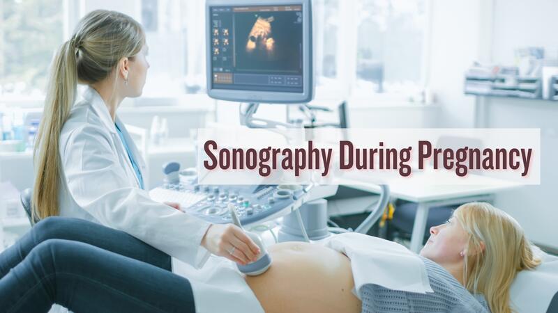
 Sonography is an ultrasound procedure used during pregnancy to track a fetus’s growth and development. Sonography during pregnancy uses sound waves to produce images of the fetus’s internal parts, evaluating advancements and checking for health issues. Here, you can explore more on this topic.
Sonography is crucial for prenatal care and can be performed at different gestation periods for different reasons. There is no inherent risk to the mother or child, and understanding the reasons and timing of sonography can help women feel more confident and ready for the changes. (1)
What Is An Ultrasonography?
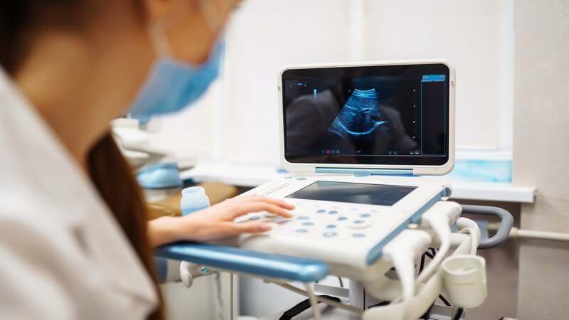
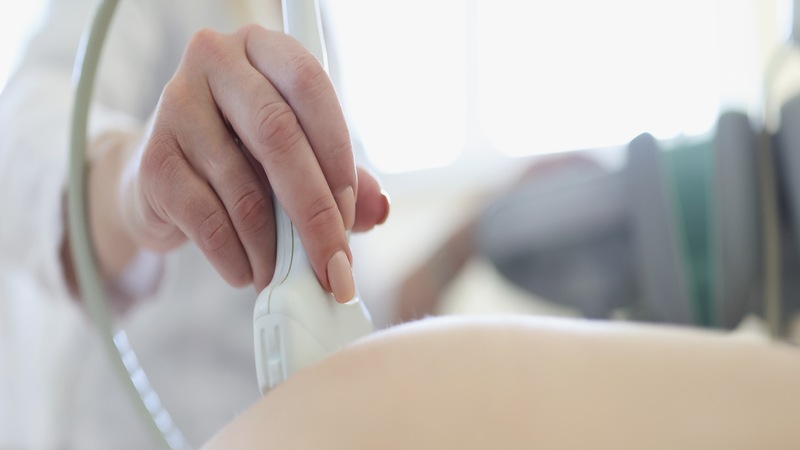
An ultrasound is a safe, non-invasive way for doctors to monitor your baby’s growth and development throughout pregnancy. It uses sound waves to create a picture of your baby, helping to spot potential problems early. While the images may not always be crystal clear, they provide valuable information, and any concerns can be followed up with additional testing if needed (2) (3).
What Is The Use Of Doing An Ultrasonography?
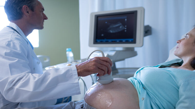
 Ultrasound, or sonography, is a painless and safe approach to checking upon your baby’s health during pregnancy. Here’s how it’s applied in each trimester (3a):
1. First Trimester (0-13 weeks)
It confirms you are pregnant and the baby is developing in the proper location. Also, it calculates how far you have progressed and estimates your due date. It lets you know if you are carrying twins or more.
2. Second Trimester (14-26 weeks)
Detailed Scan: Your baby’s inside organs, bones, and overall growth are checked at around 18-21 weeks. Gender Reveal: You will get an idea of whether it is a boy or a girl during this scan if you would like to know at this stage. Placenta and Fluid: To know where the placenta is and if there is enough amniotic fluid surrounding your baby.
3. Third Trimester (27-40 weeks)
Checking on baby’s development and position: checking to see if the baby has moved down to head position for the delivery along with checking the size. Monitoring health: monitoring that all is well, from baby development to the health of the placenta (3a) (4). Ultrasound is one of the routine tests that help in keeping your health at every stage for you and your baby.
What Can Be Detected In A Pregnancy Ultrasound?
 A pregnancy ultrasound can reveal various important information about the baby’s development and the health of the mother. Some of the things that doctors can check through this test are shown below:
- Confirms Pregnancy
- Determines Baby Due Date
- Can listen to the baby’s heart.
- View the size of your baby.
- The position of your baby can be observed.
- Any abnormalities can be confirmed.
- Fluid is present around the baby or not.
- Checking for placenta
- Checking for twins or more.
- Observe the movement of your baby (5).
How Many Types Of Ultrasound Are There?
 There are various ultrasounds conducted in pregnancies and they all have to play different purposes. Here is the general overview of types:
- Early Pregnancy/Dating Scan (8-11 weeks): Confirmation of pregnancy. Detection of fetal heartbeat. Estimation of due date.
- Nuchal Translucency Scan (11-14 weeks): Determination of the risk for chromosomal abnormalities, like Down’s syndrome.
- Early Anomaly Scan (14-18 weeks): Detection of early abnormal signs and sometimes of a baby’s sex.
- Fetal Anomaly Scan (18-24 weeks): Ultrasound check-up to ensure the baby’s organs are all in the proper place and the development is normal.
- Growth Scan (24-42 weeks): It will continue to monitor the baby’s growth, position, and liquid in the womb (6).
Your healthcare provider may request you to appear for more ultrasounds based on certain medical conditions.
How Many Ultrasounds Do You Have During Your Pregnancy?
 Most women have at least two ultrasounds throughout a healthy, typical pregnancy. Here’s a general outline of how it usually goes:
1. Early Pregnancy Scan (Dating or Booking Scan)
Done between 11 and 14 weeks to confirm the gestation period in order to see how the baby is and to give an estimate of the date of delivery. It can also include a nuchal translucency (NT) scan to check if your baby has Down’s syndrome, Edwards’ syndrome, or Patau’s syndrome.
2. Fetal Anomaly Scan (18 and 21 Weeks Mid-Pregnancy Scanning)
Scanned at 18 and 21 weeks to decide whether all the baby’s organs are developed and there is no congenital abnormality in them (7). Additional scans may be provided based on medical requirements, but these are two standard ones performed in a healthy pregnancy.
When Is The First Ultrasound During Pregnancy Conducted?
The first ultrasound, or pregnancy scan, would be between 8 and 11 weeks of gestation. The first ultrasound confirms the pregnancy, detects the heartbeat of the baby, and ensures the due date is calculated correctly. It may be scheduled between 11 and 14 weeks, depending on the provider for dating scanning to confirm the gestational age and assess early developments (8).
What Is The 20-Week Ultrasound Scan?
 The 20-week ultrasound is sometimes referred to as an anomaly scan. It occurs at around 18–22 weeks of pregnancy. It assesses the baby’s developing organs, which include the heart, brain, and kidneys, for normal formation. Also,
- The baby’s size confirms that the growth is normal.
- The position of the placenta to ensure that it is in a safe position.
- The amniotic fluid that surrounds the baby.
- You may also learn whether it’s a boy or girl if you want (9) (10).
This scan ensures that everything’s perfectly fine with the developing baby.
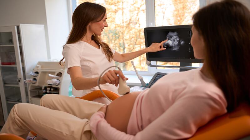
Can I See My Baby In An Ultrasound?
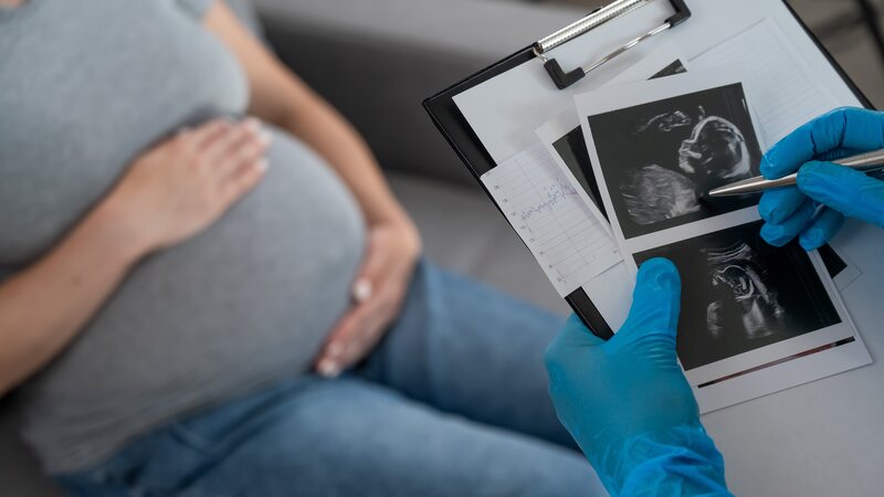
Yes, you can see your baby during an ultrasound! Here’s how it works: A sonographer performs the ultrasound and uses a handheld device called a probe to take pictures of your baby. The images will appear as black-and-white pictures on a screen. You may get a glimpse of your baby’s face, hands, legs, and other body parts. An ultrasound will help you know the age of your baby and growth, see your little one’s heartbeat, and see how your baby moves. It will also help your doctor estimate your due date (11) (12).
How To Prepare For An Ultrasound?
 Preparation for an ultrasound is simple, though it will depend on what kind of ultrasound you are getting. Here are some general tips to help you prepare for your appointment.
- If you are getting a scan during the first 12 weeks of your pregnancy, then your doctor may ask you to drink 2-3 glasses of water approximately an hour before visiting. A full bladder helps to get clearer pictures.
- You should also wear comfortable clothes
- Bring a Partner or Family Member along with you for emotional support and will also provide the opportunity for them to share in the experience of seeing the baby.
- Plan to Get There a Little Early: Arriving a little early helps you relax and attend to any paperwork (13) (14).
- If you are not sure about any information, just ask your healthcare provider for specific instructions.
What Is a 3D/4D Scan During Pregnancy?
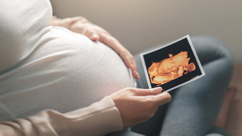 A 3D or 4D ultrasound is also an imaging procedure. It utilizes the sound waves to develop an elaborate view of the baby within the womb.
a) 3D Ultrasound
Creates a static, three-dimensional image of the baby’s surface. Such an image aids parents in viewing the features of the baby’s face and body structure in good detail.
b) 4D Ultrasound
It produces a kind of video capture that would present real-time activity, whether facial muscles or limb movement. 3D and 4D ultrasounds can identify conditions affecting the baby in particular, such as cleft palate. They are also quite handy when trying to explain abnormalities to the parents. However, when it comes to efficiency, 2D ultrasounds are quite efficient, and it is the commonly used ultrasound in checking for abnormalities in development and growth in a baby (15) (16).
Are Repeated Scans Harmful To The Baby?
No, repeated ultrasounds are not harmful to the baby if carried out according to your doctor’s recommendation. The ultrasounds also employ sound waves as opposed to radiation to generate images of the baby and there is no hazard involved in undertaking repeated ultrasounds (17) (4a). If you feel concerned, you can always ask your doctor to clarify the reasons for such repeated scans.
FAQ’s
1. In Which Month Pregnancy Sonography Is Done?
Pregnancy Sonography is performed multiple times during the nine-month pregnancy journey. The first trimester confirms the date, checks the number of babies, and ensures development. The second trimester checks the baby’s brain, spine, organs, limbs, sex, and placenta position. Finally, the third Trimester (After 30 weeks) scan ensures that your baby is growing properly and examines the placenta, ensuring that it’s not encroaching on the cervix.
2. What Are The 7 Most Common Concerns During Pregnancy?
Common concerns include morning sickness, fatigue, heartburn, backache, swelling, urinating often, mood swings due to hormonal changes while pregnant
3. Can I Drink Water Before An Ultrasound?
Yes, First-trimester pregnancy scans, which take place before 12 weeks, are most likely to require you to drink some water before attending this scan. A full bladder helps achieve a better view of the baby.
Reference
- Mayo Clinic, Fetal ultrasound – https://www.mayoclinic.org/healthy-lifestyle/pregnancy-week-by-week/in-depth/fetal-ultrasound/art-20546827
- John Hopkins, Fetal Ultrasound – https://www.hopkinsmedicine.org/health/treatment-tests-and-therapies/fetal-ultrasound
- Department of Health, Victoria State Government – https://www.betterhealth.vic.gov.au/health/healthyliving/pregnancy-tests-ultrasound#what-is-an-ultrasound
- NHS, Ultrasound scans in pregnancy – https://www.nhs.uk/pregnancy/your-pregnancy-care/ultrasound-scans/
- medically reviewed article from Cleveland Clinic – https://my.clevelandclinic.org/health/diagnostics/4995-ultrasound
- Dr Srividhya Sankaran, consultant in maternal-fetal medicine (MFM) and obstetrics, based in London. – https://www.topdoctors.co.uk/medical-articles/what-are-the-different-types-of-ultrasound-scans-in-pregnancy#
- NHS Inform, Ultrasound scans during pregnancy – https://www.nhsinform.scot/healthy-living/screening/pregnancy/ultrasound-scans-during-pregnancy/
- Lee WA, Nelson G, Grogan SP. Sonography 1st Trimester Assessment, Protocols, and Interpretation , Desert Regional Medical Center, Madigan Army Medical Center – https://www.ncbi.nlm.nih.gov/books/NBK573070/
- NHS, 20-week screening scan – https://www.nhs.uk/pregnancy/your-pregnancy-care/20-week-scan/
- Cleveland Clinic, 20-Week Ultrasound (Anatomy Scan) – https://my.clevelandclinic.org/health/diagnostics/22644-20-week-ultrasound
- Cleveland Clinic, Ultrasound in Pregnancy – https://my.clevelandclinic.org/health/diagnostics/9704-ultrasound-in-pregnancy
- March of Dimes, Ultrasound during pregnancy – https://www.marchofdimes.org/find-support/topics/pregnancy/ultrasound-during-pregnancy#
- Pregnancy, Birth And Babies, RANZCOG The Royal Australian and New Zealand College of Obstetricians and Gynaecologists, Inside Radiology (Nuchal translucency scan), Inside Radiology (18-20 week screening pregnancy ultrasound), The Royal Australian College of General Practitioners (Prenatal testing) – https://www.pregnancybirthbaby.org.au/ultrasound-scan#
- Richards DS. Obstetric ultrasound: imaging, dating, growth, and anomaly. In: Landon MB, Galan HL, Jauniaux ERM, et al, eds. Gabbe’s Obstetrics: Normal and Problem Pregnancies. 8th ed. Philadelphia, PA: Elsevier; 2021:chap 9. – https://www.ucsfhealth.org/medical-tests/ultrasound-pregnancy
- Lebit DF, Vladareanu PD. The Role of 4D Ultrasound in the Assessment of Fetal Behaviour. Maedica (Bucur). – https://pmc.ncbi.nlm.nih.gov/articles/PMC3239390/
- Robyn Horsager-Boehrer, M.D, Obstetrics and Gynecology, UT Southwestern Medical Center – https://utswmed.org/medblog/3d-4d-ultrasound/
- Newnham JP, Doherty DA, Kendall GE, Zubrick SR, Landau LL, Stanley FJ. Effects of repeated prenatal ultrasound examinations on childhood outcome up to 8 years of age: follow-up of a randomised controlled trial. Lancet. 2004 Dec 4-10;364(9450):2038-44. – https://pubmed.ncbi.nlm.nih.gov/15582061/